
What Is a Hematoma and What Types Are There?
What Is a Hematoma?
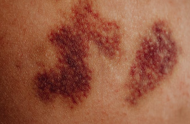
A hematoma is a collection of blood that forms outside blood vessels due to the rupture of small capillaries. It usually appears after a blow, bruise, or contusion in which the skin remains intact. Colloquially, it is known as a bruise.
Hematomas vary in size and severity depending on the affected area and the intensity of the injury.
At first, a hematoma appears red due to fresh blood under the skin. Within hours, it turns blue, purple, or even black as oxygen levels decrease. After about a week, it may take on a greenish-yellow color and eventually fade to yellow or light brown before disappearing completely.
Hematomas can occur anywhere on the body after trauma. However, when they appear without a clear cause or known injury, medical evaluation is recommended. In some cases, hematomas may be related to blood clotting disorders or the use of anticoagulant medications.
Hematoma vs. Ecchymosis: What Is the Difference?
Although the terms are often used interchangeably, hematoma and ecchymosis are not the same.
Hematoma
- Usually caused by a direct impact such as a fall, sports injury, or accident
- Size is typically larger than 5 mm
- May cause pain, swelling, and skin discoloration
- Changes color as blood coagulates and breaks down
- Usually resolves within 2 to 3 weeks
Hematomas that appear on the scalp without trauma should always be evaluated by a doctor.
Ecchymosis
- A superficial bruise that may appear without a noticeable impact
- Caused by bleeding under the skin or mucous membranes
- Measures less than 5 mm
- Often painless and resolves within 12 to 15 days
Types of Hematomas by Location
Hematomas can be classified based on the affected tissue or body area.
Subcutaneous Hematomas
These form just beneath the skin and are the most common type.
Intramuscular Hematomas
They occur within muscle tissue and can sometimes affect nearby organs. These are common in sports injuries.
Periosteal Hematomas
These develop when the injury affects a bone, causing bleeding beneath the periosteum.
Types of Hematomas by Body Region
- Epidural hematoma
- Subdural hematoma
- Intracerebral hematoma
- Scalp hematoma
- Ear hematoma
- Septal hematoma
- Subungual hematoma (under the nail)
- Pelvic fracture–related hematomas
Intracranial or Cerebral Hematoma
A cerebral or intracranial hematoma is one of the most common post-traumatic brain injuries. It involves blood accumulation within the skull, either inside the brain tissue or between the brain and the skull, usually due to ruptured blood vessels.
These hematomas may increase in size over hours or days and are classified into three main types: epidural, subdural, and intracerebral hematomas.
Acute Epidural Hematoma
This type of hematoma is most often caused by arterial bleeding due to rupture of the middle meningeal artery. It typically occurs in the temporal or parietal bone region.
Blood accumulates between the skull and the dura mater, forming a lens-shaped (biconvex) clot that compresses brain tissue.
Common Clinical Features
- A lucid interval lasting hours or days in nearly half of patients
- Rapid neurological deterioration
- Signs of increased intracranial pressure
- Pupil dilation on the affected side
- Decreased level of consciousness
Diagnosis is confirmed with a CT scan. Emergency craniotomy is required to evacuate the hematoma. Mortality rates range from 16% to 32%, but early treatment greatly improves prognosis.
Acute Subdural Hematoma
This hematoma originates from venous bleeding caused by rupture of cortical veins. It commonly affects the frontotemporal region.
The clot has a crescent (semilunar) shape and forms between the dura mater and arachnoid membrane.
Clinical Characteristics
- Immediate drowsiness or coma after injury
- Often associated with underlying brain damage
Diagnosis is confirmed by CT scan, and emergency surgery is usually required. Mortality rates can reach 40% to 50%.
Encapsulated Hematoma
In most traumatic injuries, the body gradually reabsorbs accumulated blood. However, in some cases, the hematoma becomes surrounded by fibrous tissue, forming a capsule. This is known as an encapsulated hematoma.
Physical therapy may help reduce the hematoma and prevent encapsulation.
Muscle Hematoma
Muscle hematomas are common in athletes but can affect anyone. They frequently occur in the thighs, hamstrings, quadriceps, and arms and are often associated with swelling and pain.
Frequently Asked Questions About Hematomas
How to Know If a Hematoma Is Serious
Most bruises are mild, but seek medical attention if:
- The hematoma appears on the head or neck
- It lasts longer than 15 days
- It continues to grow
- Swelling and pain worsen over time
- There is abnormal bleeding elsewhere
- Movement of a joint or limb is limited
How Is a Hematoma Treated?
Hematomas usually resolve on their own. Recommended care includes:
- Applying cold compresses during the first 24–48 hours
- Elevating the affected area
- Limiting movement
- Using pain relievers such as acetaminophen
How Long Does a Hematoma Last?
Most hematomas are visible for 2 to 3 weeks, though some may persist for months depending on location and severity.
What Is an Internal Hematoma?
An internal hematoma is caused by internal bleeding due to ruptured capillaries. The blood remains trapped within tissues and does not appear on the skin surface.
When to See a Doctor
If you have a hematoma with unusual characteristics, severe pain, neurological symptoms, or no clear cause, it is important to consult a healthcare professional for proper evaluation and treatment.